


The co-infections of Lyme disease
The Borrelia bacteria never comes alone. Ticks carry multiple bacteria and viruses with it that can also be transmitted during a bite.
Bartonella
Bartonella infections fall into the category of Tuberculosis. It is sometimes called Cat Scratch Fever because 65% of cats carry this and can transfer it to humans.
The most well known form of Bartonelliosis is Cat Scratch Fever caused by Bartonella Henselae. After someone is scratched by a cat infected by Bartonella Henselae the bacteria can infect the walls of the blood vessels. The disease can however also be transmitted by a tick infected with it after a bloodmeal on an infected cat or other animal.
Typical symptoms are pain underneath the sole of the foot, a rash in the form of stripes or a swelling in armpits or groin with pus in it.
The most well known strains are Bartonella Henselae, Bartonella Quintana and Bartonella Baciliformis. The presumption exists that the Bartonella transmitted by ticks is probably cdaused by another yet unkown strain of Bartonella. This would also explain why this variant is often not recognised by tests. Bartonella Schoenbuchii amongst others is a possible candidate.
Symptoms
The Cat Scratch Fever has an incubation period of 3 to 20 days. It often starts with one or more little lumps (2-3mm) on the skin near the scratch or bite. These quickly form into small blisters that can develop a little crust after a few days. Afterwards these little spots disappear again. Sometimes one doesn’t even notice them. After about 2 weeks the lympenodes can grow big and painful. This lymphatic infection can retain for weeks or even months (6 weeks on average) but usually disappears on its own.
With a portion of patients the disease will be accompagnied during the first few days or weeks with fever, headache, muscle and joint pains, nausea, chills and general malaise. Complaints such as shinbone pain, weight loss, sore throat and skin rash. One specific complaint is the pain underneath the soles of the feet. Bizarre neurological symptoms (sometimes), epileptic insults (sometimes), liver and spleen conditions, neuroretinitis (change of the optic nerve into tissue causing possible partial blindness), aseptic meningitis and endocarditis are also mentioned. With 2% of patients the disease can lead to meningitis with lowered consciousnes and even coma and convulsions. With people who have lowered immunity the disease usually runs a more severe course. In this case one can first find lumps and bleeds in the skin, spleen and liver. They can be accompanied by fever and general malaise and sometimes even lead to death.
Treatment of Bartonella is not simple. A few antibiotics have proven effective against Cat Scratch Fever. Gentamicine, Rifampicine and Ciprofloxacine are mentioned. The disease can heal on its own provided the patient doesn’t have any other tickborne diseases. The disease is difficult to diagnose through bloodwork. As far as testing is concerned, we see the same problems we do with Lyme-Borreliose.
Babesia
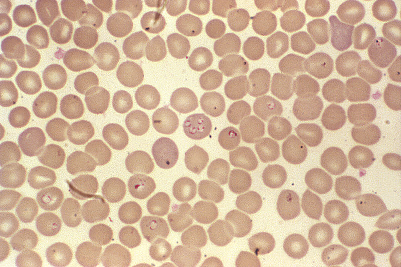
Babesiosis, tick fever or piroplasmosis, is an infection that is caused by the parasite babesia. There are multiple strains known of this parasite. Babesia is a protozoa comparable to the causing agent of malaria. Babesiosis just like Lyme-borreliosis are transmitted by ticks. Infection with this parasite in animals is the most common but it can also infect humans. After a tick bite, the incubation period is 1 to 3 weeks, 6 weeks at most. After a blood transfusion this can even be 9 weeks.
Babesia lives in red bloodcells and destroys them. This causes fever, headaches and muscle pain. The breaking down of bloodcells can cause anemia. For healthy people recovery normally occurs without treatment within 2 to 3 weeks. For people with diminished immunity, like people without a spleen, HIV-infected people, corticosteroid-users or people who have been infected by a borrelia infection simultaneously or the elderly the infection can lead to serious problems and even be fatal. Until recently this ailment was limited in Europe to warm and to a lesser extent moderate areas. In the USA this is becoming epidemic and is mainly spread through bloodbanks.
Symptoms
Serious malaise and fatigue (exhaustion), severe day and night sweats, high fever and chills, weakness, weight loss, nausea, vomitting, diarrhea, cough, shortness of breath, sever headache, muscle pains, stiff neck and back, dizziness, anemia, dark urine (or blood in urine) and severe neuro psychiatric symptoms.
Ehrlichia (anaplasma)
The illness Ehrlichiosis or Anaplasma is caused by a bacteria. Ehrlichia spp. Can by transmitted by ticks. This bacteria is in the Netherlands long known by veterinarians as transmitter of disease in small herbivores, cows and horses. Then it concerns Ehrlichia phagocytophilia and Ehrlichia equine. In humans two distinct species of erkichia can occur: Humane Monocytarian Ehrlichiosis (HME) and Humane Granular Ehrlichiosis (HGE). HME which is caused by the bacteria Ehrlichia Chaffeensis was first found in humans in the early eighties in the USA. In Europe we haven’t encountered this disease yet. HGE has been found in Europe since a few years and recently (1999) also in The Netherlands. Because it is still unknown which Ehrlichia species is responsible for HGE in humans, the agent is labeled Ehrlichia spp. The bacteria settles in white bloood cells and diminishes the resistance of humans.
Symptoms
Symptoms usually appear after 1 to 2 weeks following a tick bite. They include: severe headache, fever, muscle aches and spasms, joint pains, stomach aches, lesser appetite and swollen lymp nodes. De fever lasts anything from 2 tot 11 days. Othe less common symptoms include: nausea, abdominal pain, diarrhea and coughs. Also being reported are: dizzyness, vomitting, confusion, rashes, clotting disorders usually accompagnied with trombopenia (lowerd blood platelets), anemia, leucopenia (lowered white bloodcells), higher liver enzymes and splenomegalia (larger spleen), central nervous system impairment and bizarre neurological symptoms.
Ehrlichiosis can be treated very well with antibiotics. In case of Lyme-Borreliosis this infection needs to be treated similtaneously.
FSME (Fruh Sommer Meningo Encephalitis)
FSME is also known as TBE (Tick Borne Encephalitis) or TBD (Tick Born Disease) and is caused by a virus. It is an inflammation of the brain or the cerebral membrane.
Symptoms
The disease has 2 stages. The first stage begins with flue like symptoms, fever and headache. This lasts for about 5 to 10 days. After that period there is a symptom free period of 4 to 10 days. As with other infections here mentioned, the majority of the disease runs its course unnoticed. Sometimes however, one can experience a slight flu like feeling.
The second stage is the inflammation of the brain and the cerebral membrane. By a portion of the infected people this second stage occurs. Of those that enter second stage, 3 to 5% will have lasting neurological conditions (paralysis, deafness, headaches). A small percentage (1-2%) even die of FSME. About half of the cases retain lasting damage from this disease.
FSME is spread all over Europe, like Eastern Europe, Scandinavia, Central Europe, Southern Germany and France.
Since the FSME virus is located in the saliva gland of the tick, the virus will be transmitted to humans right at the start of the sucking process.
With FSME, in contrary to Lyme disease it is possible to inocculate yourself beforehand. This treatment requires 3 injections with FSMEimmun® that are given spread over the course of a year and then provide protection for 3 years afterwards. Every 3 years you will need to get a new injection. Because the disease is caused by a virus, a treeatment with antibiotics isn’t useful.
Rocky Mountain Spotted Fever (RMSF)
RMSF is an infectious disease caused by the bacteria Rickettsia rickettsii. People can get infected with this bacteria through a tick bite of a tick carrying the bacteria and can get really ill from it. RMSF is a serious infectious disease which usually requires a hospital addmittance. Despite treatment, about 3 to 5% of patients don’t survive this disease.
Symptoms
Usually the disease manifests within 2 weeks after a tick bite. On the spot the bite occurred, a black or dark crust forms and a rash spreads from the feet or hands quickly all over the body. These symptoms involve a suddenly rising high fever, fatigue, sever muscle pains and headaches or other pains. During physical examination the spleen and the liver often turn out to be enlarged. A complication that can occur with RMSF is gangreen (dying off of tissue due to lesser blood supply) in fingers and toes. If this is not treated in time, surgical removing of those affected parts can be necessary.
An other possible complication is parotitis, an infection of the ear gland. In sever cases pneumonia or kidney failure can occur.
Treatment
RMSF is treatable with antibiotics like tetracycline or doxycycline. When treatment is started early, prognosis is good.
Fièvre Boutonneuse (FB)
In the Mediterranean countries FB is very frequent. This disease is caused by the bacteria Rickettsia Connorii. In our regions this disease is sometimes imported from the Mediterranean. Humans can also contract the disease by removing ticks from pets like the dog. Very recently a study found that 20% of the studied Dutch ticks have Ricjettsia Helvetica.
Symptoms
On the spot of the tick bite a small sore (tâche noir or black spot) forms. The lympf nodes are often also swollen. Usually smaller blood vessels get infected too that can then get clothered and/or cause small internal bleedings. Headaches, muscle pains and joint pains can occur often together with lowered blood pressure and sometimes with neurological disorders and disorders of the renal function. After 4 to 5 days red spots emerge usually on the hand palms or footh soles. The fever that occurs after 5 to 7 days can be accompagnied by cold chills, is moderately high or high and disappears quickly after antibiotic treatment. The disease is usually not very severe and can heal naturally.
Chlamydia pneumoniae (CPN)
Chlamydia pneumoniae is an intercellar bacteria that knows 3 life forms just as Lyme does. It can itself not make energy and hijacks the energy of it’s hosts, humans. This often results in extreme exhaustion but it can also result in burning pains in the chest, the bones or the muscles. Chlamydia pneumoniae is contagious, airborn and all around us. Through the airways it enters the lungs and infects the lung tissue. CPN can be spread from the lungs to other body parts. There it can grow in the cells of its host. CPN steals energy from the cells to grow (ATP). It can also spread further to the connective tissues, nerve tissues, brain, muscles, macrophages and others. It infects the enothelial cells, cells that are present in vains and the lypmhatic system.
Symptoms
Pneumonia, meningo-encephalitis, arthritis, myocarditis, connective tissue pains (fybromyalgia), asthma, infections of the airways, bronchitis, MS-like complaints, Guillian Barre, Alzheimer, atherosclerosis, shortness of breath and continued slime production.
CPN can have a more serious progress in immunocomprised patients. Once embedded into the cells and tissues, CPN can become chronic. Antibiotics have a hard time treating CPN. Its has a life cycle of 3 phases, can retain intra cellular and can become resistant by gen-transfer.
Q Fever
Q fever is caused by the Coxiella Burnetii bacteria. The contammination by humans probably goes through the lungs but we can’t overlook ticks as causing agents here either. Comtamination through the stomach and bowel system by consuming raw milk or derivate products (like butter, cheese under 6 weeks) or by eating meat that hasn’t been heated properly are also possible.
Humans can not only be infected by cows, sheep and goats but also by dogs, cats and birds. Infection can occur by breathing contaminated dust from stables, pastures, rough wool and animal skins, from direct contact with infected animals and by consuming contaminated raw milk of not sufficiantly heated contaminated meat. The risk of infection is highest when young cattle is being born. Birth residue like afterbirth and amniotic fluid when they are not disposed of adequately or in time can be a risk factor. Only one bacteria is enough to get infected. The incubation period of Q fever is 9 to 40 days.
Animals can dispose of large quantities of C. Burnetii through faeces, urine, milk, placenta and amniotic fluid. Inhalation of contaminated dust from stables, pastures, rough wool, hides, clothing, etc. can cause infecftion.
The bacteria is prevalent all over the world. Previously the bacteria was catalogued under Rickettsia.
Symptoms
Flu like symptoms with sudden high fever with possible complaints like chest pain and possibly also hearth problems. Further more heavy headaches, chills, sweats, muscle pain, nausea, vomitting and diahrea are mentioned. Some patients also have conditions of the airways and a dry cough. Q Fever can sometimes also cause bacterial arthritis. About 30% of patients with a chronic form have a high blood sediment and a lowered hearth rate.
A big portion of residents of The Netherlands (asymptomatic) are infected in early childhood and develops antibodies. This can be easily explained by the big cattle stock in The Netherlands where C. Burnetii under favorable climatological circumstances can be spread (through the air) over large areas.
The treatment of an acute infection usually involves an antibiotic course for 2 to 3 weeks. Q Fever often heals on its own. Tetracyclides are believed to cut down the time the fever lasts in half if administered within the first 3 days of being ill. The treatment advise is to administer doxycycline 100mg twive a day for 15 to 21 days. The optimal treatment of chronic Q Fever and Q Fever endocarditis isn’t set but usually consists of a combination of tetracycline with rifampicine or trimethroprim-sulfamethoxazole for a minimum duration of 2 to 3 years.
Coxsackie virus
The coxsackie virus is an enterovirus. Enteroviruses are very small viruses in the intestinals and the stool. There are 2 main groups of coxsackie viruses: type A and type B. Under these main groups fall yet more smaller groups of different strains of coxsackie viruses. The virus is named after the place Coxsackie in the state of New York where it was first discovered in a patient.
Coxsackie viruses can result in all sorts of symptoms varying from only a fever to sore throat, diarhea, vomitting, rash, muscle pains, liver inflammation and inflammation of the heart sack. With babies a coxsackie viral infection can be very serious resulting in for example meningitis, blood poisoning or pneumonia. Coxsackie virus is also the causing agent for hand foot and mouth disease.
Diagnosis is set through lab work where faeces, muccus from the throat, urine or brain and spinal fluid are tested.
The treatment is aimed at combatting the symptoms because the virus itself cannot be treated.
One of the clinical pictures caused by the virus is called Bornoholm disease. In this syndrome the pleura is infected (pleuritis) which results in sever chest pains that can come in seizures.
Yersinia
This bacteria can infect bowels and airways. The bacteria from the genus Yersinia are gram-negative enterobacteria. From the 17 described species there are 3 know to be human pathogens: Yersinia Pestis, Yersinia Pseudotuberculosis and Yersinia Enterocolitica. Formerly they were know as Pasteurella Pestis named after Louis Pasteur. In 1940 the name was changed to Yersinia after Alexandre Yersin who in 1894 first described the bacteria Yersinia Pestis (causing agent of the plague) as a contributor at the famous Pasteur Institute.
Infection arises after ingestion of the bacteria through contaminated food or beverages. Transmission is also possible after a blood transfusion. The most common infector is raw or not fully heated through pork. Other foods that have been connected to causing Yersinia are non pasteurized milk and milkproducts, meat, tofy, oysters and fish. Raw vegetables can carryt the bacteria on their surfaces. Pasteurized milk and milk products can get infected too because the bacteria survives in cooled environments. The most important animal reservoires for this bacteria are pigs, dogs, sheep, rodents and cats. The transmission between humans and feco-oral transmission are not proven and are deemed unlikely.
An infection with Yersinia Enterocolitica results in diarhea, low fever and abdominal pain as most significant symptoms. Vomitting occurs in about 15-40% of cases. Enterocolitis is the most common manifestation of an Y. Enterocolitica infection.
It mainly occurs within young children of around 2 yeras old. The incubation period is 4 to 6 days and the earliest symptoms are apathy, headache and lack of appetite. Afterwards the patient will develop a watery diahrea, fever and abdominal pain. These symptoms can last from 1 to 3 weeks but enterocolitis usually has a selflimiting course. Especially in young children but adults can develop in rare cases serious complications like acute appendicitis, ulceratis and inflammation of the small intestine, peritonitis, meningitis and cholangitis.
In older children and young adults symptoms are mainly acute pseudo-appendicitis, adenitis and terminal ileitis. These conditions have alongside nausea, vommiting and diahrea also the possible symptoms of fever, ebdominal pain near the right under quadrant and leucocystosis.
Almost 10% of patients with non diagnosed symptomatic Y. Enterocolitica infection turned out to have had a laparaptomy due to the presumption of appendicitis. More rare sequelae of an infection are possible. A form of reactive arthritis can occur in several joints that can typically last from 1 to 2 weeks after the intestinal infection with Y. Enterocolitica and up to 4 months. Even rarer complications include myocarditis and glomerulonefritis.
Septicemia is a rare complication where by the bacteria spread out of the gastro)intestinal system into the blood stream. This usually occurs when there are underlying causes or diseases like diabtis mellitus, alcoholism, malnutrition, immunodeficiency or a blood disease. This then becomes a life threatening situation that requires emergency medical attention. Metastatic infections after Y. Enterocolitica sepsis are possible: abcesses in liver, kidney’s, spleen or lungs. Skin infections and leasions, pneumonia, meningitis, endocarditis and osteomyelitis can occur.
Mycoplasma
Mycoplasma is a strain of bacteria. It is contagious thorugh coughing and direct contact. For people with deprived immunity the consequences of a mycoplasma infection can be severe.
Symptoms
Fatigue, fever, joint complaints, muscle aches, sleeplesness, head aches, anxieties and emotional lability, memory and concentration problems and confusion.
As treatment antibiotics can be used.
The bacteria don’t have cell walls. This makes them very vulnerable. There are more than 100 mycoplasma species known. They cause disease in animals (mycoplasmosis). In plants they are called fytoplasmas. Because they don’t have cell walls they can’t grow very big. They are the smallest bacteria that are currently known. The colonies of most Mycoplasma species look like a sunny side up egg. Some species look like a drop of water. This sunny side up effect is caused by their tendency to grow in the agar which leads to a thickening in the middle of the colony. They are so tiny that scientists when they were discovered thought they had found a new sort of virus. The colonies of the Mycoplasma species are so small they can’t be detected with a light microscope.
Mycoplasmata colonize muccus membranes of the airways and the tractus urogenitalis. The bottle shaped cells of M. Pneumoniae have a terminal tip structure responsible for adhesion to cell membranes. The bacteria can enter leucocytes and macrophages. When the infection concerns the trachea, bronchiola and peribronchial tissues, one can see purulent exsudate in the airways consisting of mainly polymorfnuclear leucocytes. There is infiltration of the bronchial walls and bronchiola with monocytes, especially plasma cells. As a result of the infection there is a loss of cilia and complete desquamation of cilian airway epithelium.
This page is also available in:
 Nederlands


